
Published in Nature Communications: SoPh ZJU Team Reveals the Molecular Basis of Subtype Selectivity in Kinin Receptor Antagonists
2026-05-27 | 药学院英文网
The kallikrein–kinin system, or KKS, is an important signaling network involved in vasodilation, inflammation and pain. Two G protein-coupled receptors within this system—the bradykinin type 1 receptor, or B1R, and the bradykinin type 2 receptor, or B2R—have attracted considerable interest as potential therapeutic targets. Antagonists targeting these receptors have shown therapeutic potential in conditions including angioedema, pancreatitis and asthma. However, the molecular mechanisms that enable antagonists to distinguish between the two receptor subtypes have remained incompletely understood.
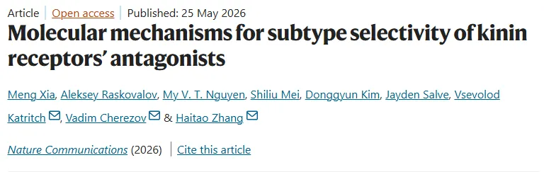
On May 25, a collaborative research team from the School of Pharmacy, Zhejiang University, or SoPh ZJU, and the University of Southern California published a research article titled Molecular mechanisms for subtype selectivity of kinin receptors’ antagonists in Nature Communications. The study was led by corresponding authors Haitao Zhang of SoPh ZJU and Vsevolod Katritch and Vadim Cherezov of the University of Southern California.
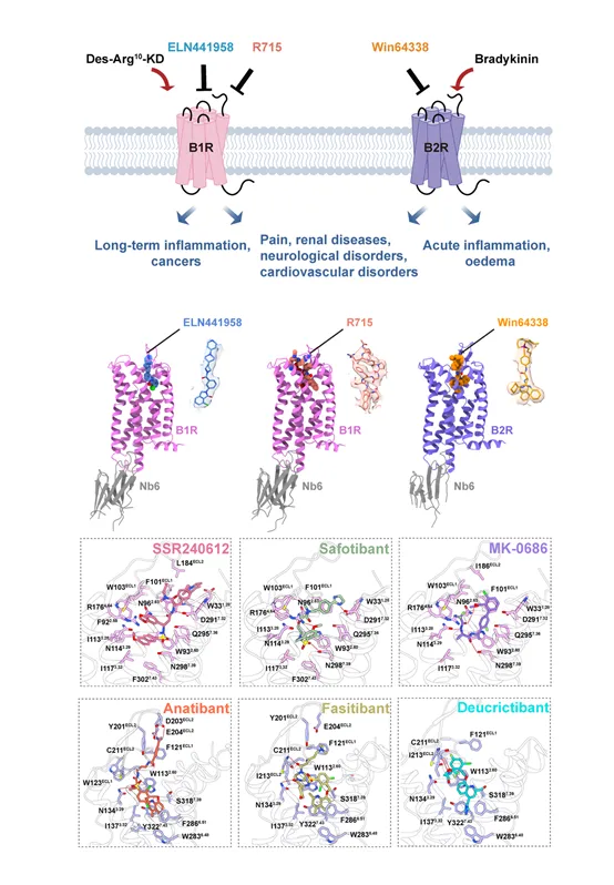
Using cryo-electron microscopy, together with functional assays and molecular docking, the researchers investigated antagonist recognition and subtype selectivity and determined three receptor–antagonist complex structures: B1R bound to the peptide antagonist R715, B1R bound to the small-molecule antagonist ELN441958, and B2R bound to the small-molecule antagonist Win64338. These structures provide detailed views of how peptide and small-molecule antagonists interact with the ligand-binding pockets of the two kinin receptor subtypes.
Comparative structural analysis, sequence analysis and site-directed mutagenesis identified three non-conserved residues at the bottom of the ligand-binding pocket as major determinants of small-molecule antagonist subtype selectivity. The corresponding residues are Y266, N298 and F302 in B1R, and F286, S318 and Y322 in B2R. Differences in the size and chemical properties of these residues reshape the lower region of the binding pocket, thereby influencing which antagonist structures can be accommodated by each receptor.
The bottom of the B1R binding pocket is comparatively compact and favors smaller hydrophobic groups, whereas the corresponding region in B2R is more spacious and can accommodate larger hydrophobic moieties. Among these residues, S318 in B2R was shown to play a particularly important role in the B2R selectivity of Win64338. Sequence and conformational differences in the second extracellular loop, or ECL2, may also contribute to antagonist selectivity.
The study also uncovered a shared mechanism by which antagonists inhibit B1R and B2R. By occupying the orthosteric ligand-binding pocket and forming hydrophobic and polar interactions with the receptor, the antagonists restrict conformational changes in the conserved toggle-switch tryptophan residue, W6.48. This prevents the structural rearrangements required for receptor activation and consequently suppresses downstream G protein signaling.
Together, the findings provide an atomic-level structural framework for understanding how peptide and small-molecule antagonists recognize B1R and B2R differently. The work may facilitate the structure-guided design and optimization of subtype-selective kinin receptor antagonists, with potential implications for future research into treatments for inflammatory disorders and pain.
Meng Xia and Shiliu Mei of SoPh ZJU, together with Aleksey Raskovalov and My V. T. Nguyen of the University of Southern California, contributed equally to the study as co-first authors. Haitao Zhang, Vsevolod Katritch and Vadim Cherezov are the corresponding authors.
NEWS
-
28
2026.07
-
17
2026.07
-
03
2026.07
-
08
2026.06
-
27
2026.05
-
15
2026.04